

West Nile
<< Back to Cases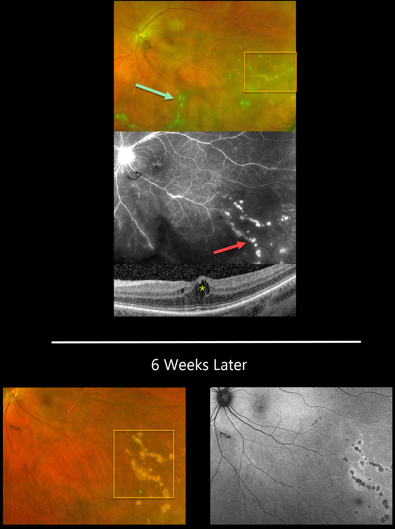
WEST NILE VIRUS
Lisa Faia
Originally posted on @retina.rocks June 28, 2023
This 51YO female presented with a recent history of floaters in her left eye. There was no significant past ocular or medical history. Vision was 20/30 OS, and the right eye was normal.
Optos RG imaging shows clumps of vitreous floaters over the posterior pole, extending into the inferior periphery. A few retinal hemorrhages are noted in the temporal midperiphery, and faint, small, multifocal atrophic scars are seen inferotemporally.
Fluorescein angiography better shows these multifocal scars with late staining. There is late optic nerve leakage and variable focal staining of the retinal vessels. OCT scanning shows mild cystoid edema.
We were concerned for possible herpes viral retinitis, and she was started on valtrex, had an anterior chamber tap, and a uveitis workup. The tap came back negative for herpes viruses. West Nile titers were ordered, and these came back positive for IgG (2.48) and low but detectable for IgM (0.10). Valtrex was stopped, and Durezol QID OS was started with a slow taper.
When she returned 6 weeks later, the vitreous was clear, and linear clumps of atrophic scars followed the retinal vessels. The scars were hyper-autofluorescent with small rims of increased autofluorescence.
Learning Points:
West Nile virus is an RNA virus transmitted by the bite of a mosquito that has fed on an infected bird. Although most infections are asymptomatic, about 20% will develop fever, weakness, headache, myalgia, nausea, and skin rash.
Multifocal chorioretinitis with or without vitreous inflammation is the most common ocular finding, occurring in about 80% of cases. Acutely, there are usually about 10-50 deep, 200-1000-micron yellow-white, scattered or radiating, curvilinear, clustered lesions that are relatively depigmented upon resolution.
Treatment of the primary viral infection is supportive. See a great review on this topic by Garg and Jampol (Surv Ophthalmology 2005;50:3-13).
WEST NILE VIRUS
Originally posted on @retina.rocks October 8, 2020
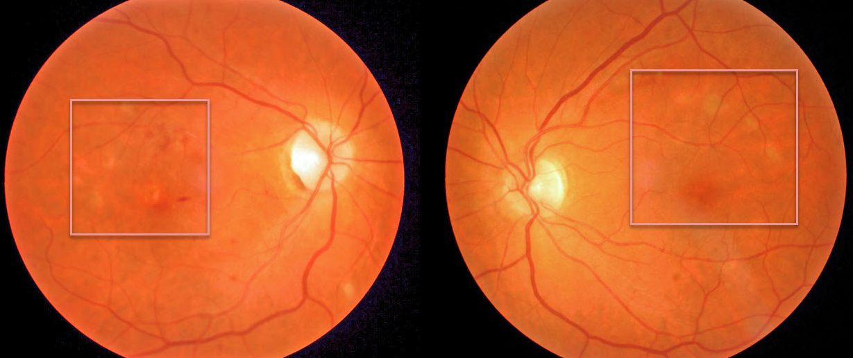
Our patient presented with macular pigmentary changes, which were most pronounced in the right eye. Vision was counting fingers OD and normal OS.
On further questioning, they gave a history of vision loss following West Nile encephalitis 6 months earlier. Although we can’t say for sure, these findings are most likely the sequelae of resolved West Nile chorioretinitis.
Learning Points:
West Nile virus is an RNA virus transmitted by the bite of a mosquito that has fed on an infected bird. Only 1 in 150 of infected persons experience encephalitis, and far fewer will have ocular effects.
Chorioretinitis in a linear pattern is the most common ocular finding, although retinal vasculitis and optic nerve swelling can also occur. There is no proven treatment for West Nile virus infection.
See a great review on this topic by Garg and Jampol (Surv Ophthalmology 2005;50:3-13).


