

White Dot Fovea
<< Back to Cases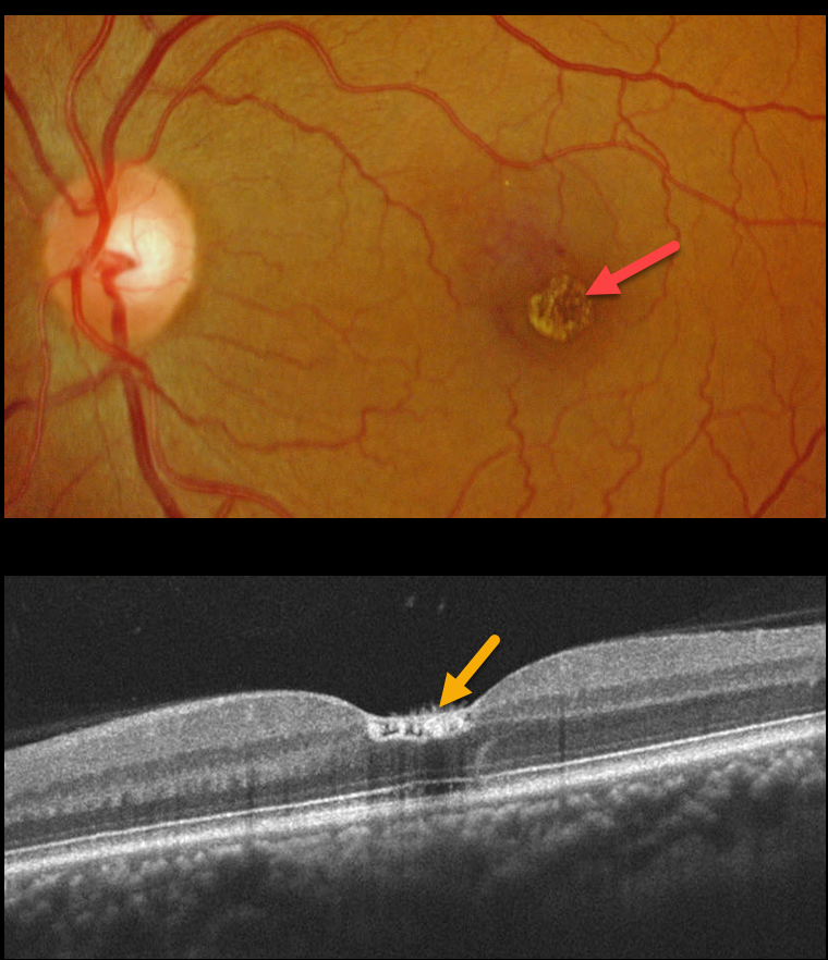
WHITE DOT FOVEA
Originally posted on @retina.rocks April 19, 2022
This 47YO male has 20/30 vision acuity and discrete unilateral inner foveal white dots. He was recently hospitalized with double pneumonia and was significantly immunocompromised.
OCT shows foveal hyperreflective deposits extending from the internal limiting membrane (ILM) to the outer plexiform layer. Some hyperreflective material is also noted within the ILM. The right macula was normal. We felt this most likely represented the white dot fovea.
Learning Points:
Initially described by Yokotsuka et al (AJO 1997;123:76-83), white dot fovea appears as usually bilateral tiny foveal white dots distributed diffusely on the inner fovea or along its margin. Histologically, it remains unclear what the white dots represent, but the configuration may suggest Müller cell end feet.


