

White Without Pressure (WWP) + Dark Without Pressure (DWP)
<< Back to Cases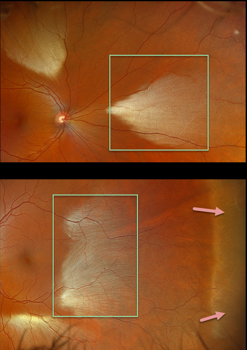
MYELINATED NERVE FIBER LAYER + WWP
Originally posted on @retina.rocks May 18, 2023
This healthy 35YO male was referred for asymptomatic fundoscopic changes. Vision was 20/20 OU.
Opto imaging shows large multifocal patches of myelinated nerve fiber layer (NFL) in both eyes. Temporal white without pressure (WWP) is also noted in his left eye.
Learning Points:
Myelinated NFL is a relatively uncommon, unilateral or bilateral, usually isolated finding present at birth. The myelinated NFL is often connected to the optic nerve, but not necessarily as in this case. Most patients are asymptomatic, although some may have a corresponding relative scotoma. The myelination can progress in about 10% of cases and can also resolve following retinal ischemic events.
Myelinated NFL occurs when retinal nerve fibers develop a myelin sheath, which usually stops posterior to the lamina cribrosa. The myelination may represent an oligodendrocytic choristoma (see Rao et al Retina 2019;39:1125-1132).
WWP is a relatively common benign peripheral finding, most often seen in darkly pigmented individuals. Typically, there are bilateral peripheral geographic areas of whitish retinal discoloration. WWP gets its name because the area appears white without indentation, unlike scleral depression (white with pressure). It can sometimes be confused with retinal breaks or detachment.
The exact cause of this phenomenon is unknown. Historically, it was felt to be due to a benign vitreoretinal interface change, but more recent OCT findings show increased reflectivity in the outer retina (see Diaz et al, Retina 2014;34:1020-1021).
RHEGMATOGENOUS RETINAL DETACHMENT
Originally posted on @retina.rocks April 15, 2022
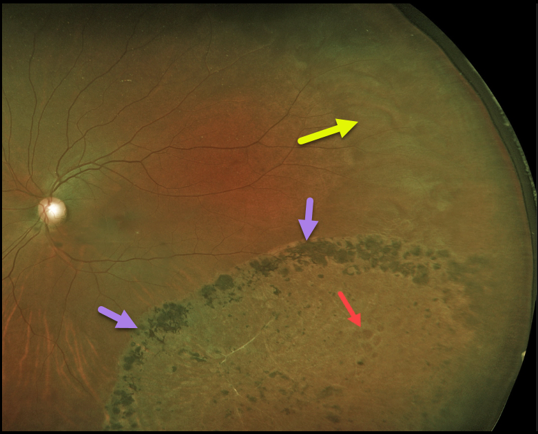
Years earlier, this 44YO female underwent laser demarcation of an asymptomatic inferotemporal rhegmatogenous retinal detachment (RRD). The prior detachment remains well demarcated, but the detachment itself has completely flattened.
Some of the causative lattice-related atrophic holes are seen within the area of prior detachment. White without pressure is also noted in the temporal periphery.
Learning Points:
Demarcation, rather than reattachment, of the retina is a valid option for select cases of RRD. When demarcating these detachments, it is critical that the retinopexy completely surrounds the subretinal fluid and that treatment extends to the ora; otherwise, the detachment can spread through the untreated retina.
Patients need to be followed postoperatively for life since the detachment can occasionally progress through the prior retinopexy.
Interestingly, we have seen that some of these detachments, as occurred in this case, spontaneously flatten following demarcation.
See Vrabec and Baumal, Ophthalmology 2000;107:1063-1067 for an excellent review regarding demarcation laser photocoagulation for selected macula-sparing retinal detachments.
DARK WITHOUT PRESSURE + CHRPE
Originally posted on @retina.rocks April 7, 2022
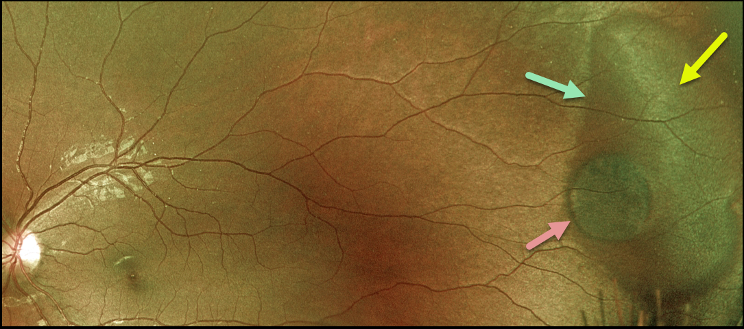
This 22YO female presented with a large temporal patch of dark without pressure (DWP), white without pressure (WWP), and a small round patch of congenital hypertrophy of the RPE (CHRPE). None of these findings is of concern, but we thought it was cool that all of them are associated with each other at this single location.
Learning Points:
WWP and DWP are relatively common benign peripheral findings most commonly found in darkly pigmented individuals. Typically, there are bilateral peripheral geographic areas of whitish (WWP) or dark (DWP) retinal discoloration.
WWP gets its name because the area is white without indentation, such as during scleral depression (white with pressure). It can sometimes be confused with retinal breaks or detachment.
The exact cause of this phenomenon is unknown. Historically, it was thought to be due to a benign vitreoretinal interface change, but more recent OCT findings show increased (WWP) or decreased (DWP) reflectivity in the outer retina (see Diaz et al., Retina 2014;34:1020-1021).
Although CHRPE lesions can enlarge with time (see Shields et al, Ophthalmology 2003;110:1968-1973), they carry virtually no malignant potential. Histologic RPE hypertrophy presents in several ways.
The typical isolated CHPRE lesions (this lesion) and bear tracks are not associated with intestinal polyps or cancer, and thus require no systemic evaluation. Only the multiple, small comet-shaped lesions are part of the familial polyposis spectrum.
DARK WITHOUT PRESSURE
Originally posted on @retina.rocks August 31, 2021
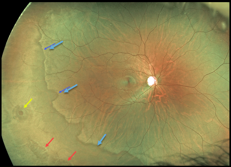
This 22YO male presented with bilateral dark without pressure, lattice lesions, and atrophic holes. He is asymptomatic, and prophylactic treatment was not recommended.
Learning Points:
White without pressure (WWP) and dark without pressure (DWP) are relatively common benign peripheral findings most commonly found in darkly pigmented individuals. Typically, there are bilateral peripheral geographic areas of whitish (WWP) or dark (DWP) retinal discoloration.
WWP gets its name because the area appears white without indentation, unlike scleral depression (white with pressure). It can sometimes be confused with retinal breaks or detachment. The exact cause of this phenomenon is unknown.
Historically, it was felt to be due to a benign vitreoretinal interface change, but more recent OCT findings show increased (WWP) or decreased (DWP) reflectivity in the outer retina (see Diaz et al, Retina 2014;34:1020-1021).
WHITE WITHOUT PRESSURE
Originally posted on @retina.rocks May 21, 2021
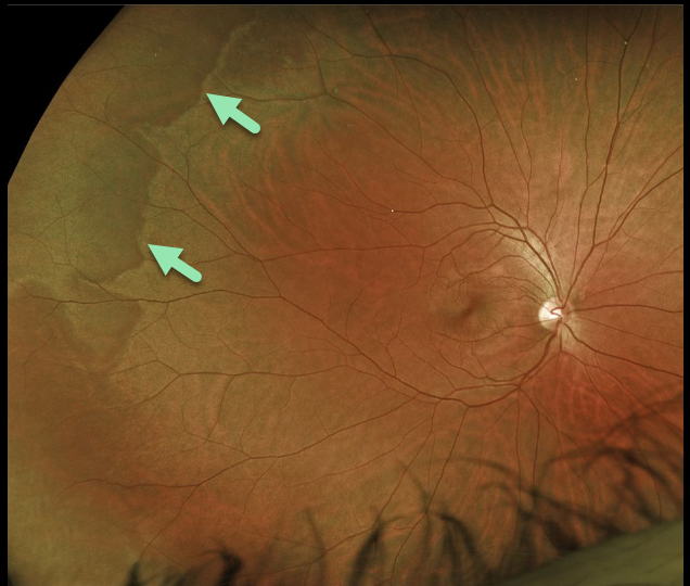
This patient’s fundus shows areas of white without pressure. Typically, there are bilateral peripheral geographic areas of whitish retinal discoloration.
Learning Points:
White without pressure (WWP) is a common benign peripheral finding, most often seen in darkly pigmented individuals. WWP gets its name because the area appears white without indentation, unlike scleral depression (white with pressure).
It can sometimes be confused with retinal breaks or detachment. The exact cause of this phenomenon is unknown. Historically, it was thought to be due to a change at the vitreoretinal interface, but more recent OCT findings show increased reflectivity in the outer retina (see Diaz et al, Retina 2014;34:1020-1021).
RETINAL BREAK
Originally posted on @retina.rocks January 13, 2021
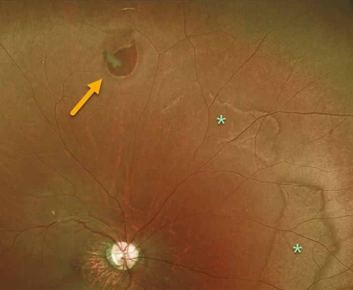
This is a 19YO patient who presented with an asymptomatic retinal break with prominent posterior white without pressure.
Learning Points:
A retinal break is a full-thickness retinal defect. Breaks are classified as atrophic holes, operculated holes, tractional (horseshoe) tears, and retinal dialysis
(break at the ora serrata).
In general, asymptomatic retinal breaks are observed since they are at low risk of causing retinal detachment. Symptomatic retinal tears and retinal dialysis are at the highest risk for causing a detachment and are virtually always treated.


