

X-Linked and Non-X-Linked Retinoschisis
<< Back to Cases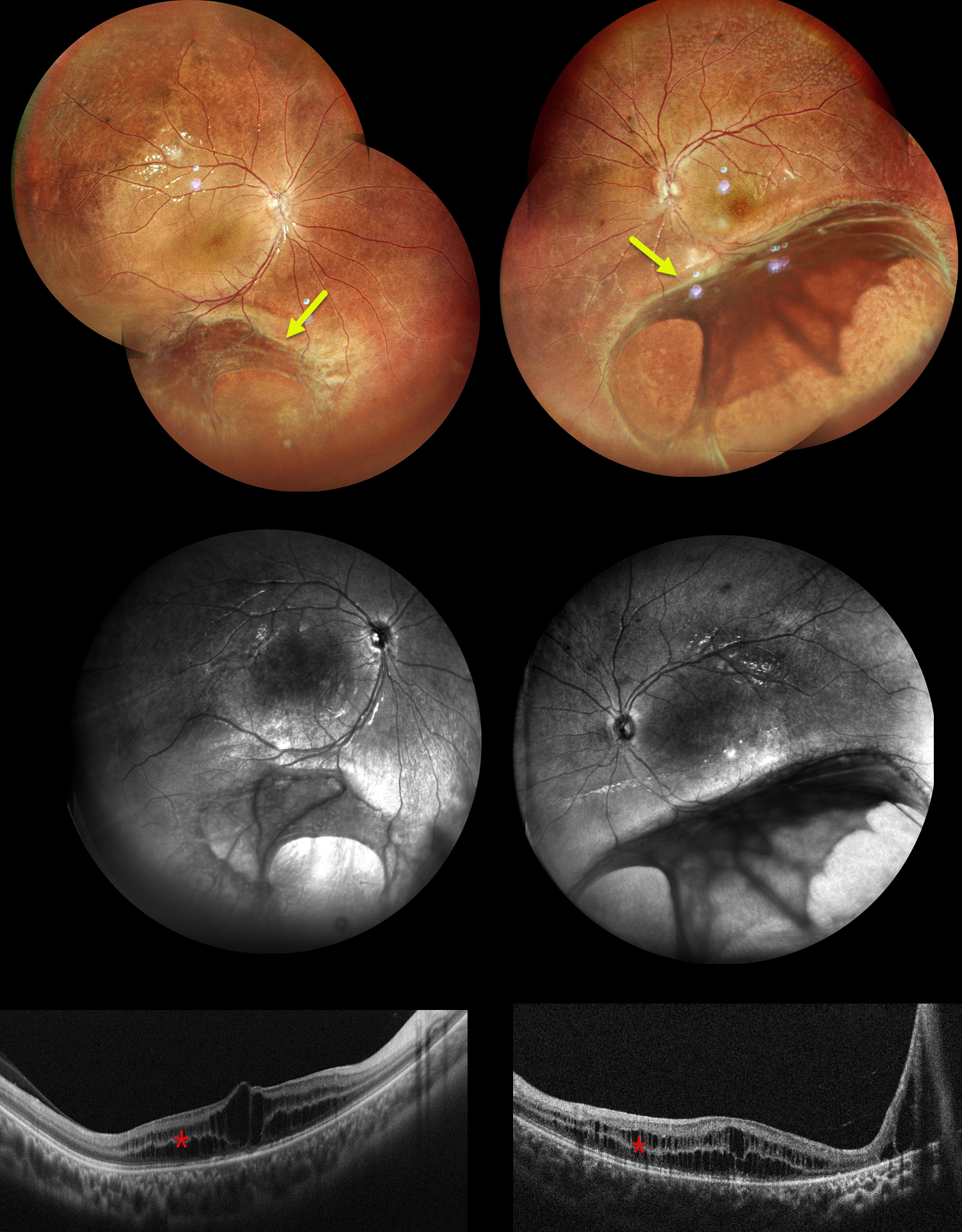
X-LINKED RETINOSCHISIS
Shraddha Raj Shrivastava, Akansha Sharma and Manish Nagpal
Originally posted on @retina.rocks August 19, 2026
This 9YO boy presented with decreased vision in each eye since early childhood. Vision was 20/80 OU.
Pseudocolor SLO imaging shows blunted foveal reflexes due to inner and outer nuclear layer schisis on OCT. Widefield imaging shows extension of the schisis cavities into the inferotemporal periphery, involving the inner retina with vitreous veils. These changes are best seen in the infrared images. Our patient’s 13YO brother was examined the same day and had similar fundus findings.
Both brothers were prescribed topical carbonic anhydrase inhibitors (dorzolamide) and asked to follow up in 3 months. Genetic testing was recommended.
Learning Points:
Sex-linked retinoschisis is caused by a hemizygous mutation in the RS1 gene, which is located on the X chromosome and encodes for retinoschisin, a protein secreted by photoreceptors that is involved in intercellular adhesion and likely in retinal cellular organization (Heymann et al, Progress Retinal Eye Research 2023;95:101147). Although usually transmitted as an X-linked recessive disorder in males with an incidence of 1 in 15,000 to 30,000, it can also sometimes affect females.
The characteristic features include “spoke-wheel” foveoschisis and peripheral schisis with vitreous veils. Over time, the macular schisis flattens with secondary atrophy, and vision usually levels off at about 20/100. Patients can develop spontaneous vitreous hemorrhage and rhegmatogenous retinal detachment from peripheral schisis. Although there is no specific treatment, topical carbonic anhydrase inhibitors may decrease the foveal thickness and possibly help minimize foveal atrophy (see Andreuzzi et al Retina 2017;37:1555-1561).
STELLATE NON-HEREDITARY IDIOPATHIC FOVEOMACULAR RETINOSCHISIS
Originally posted on @retina.rocks May 7, 2025
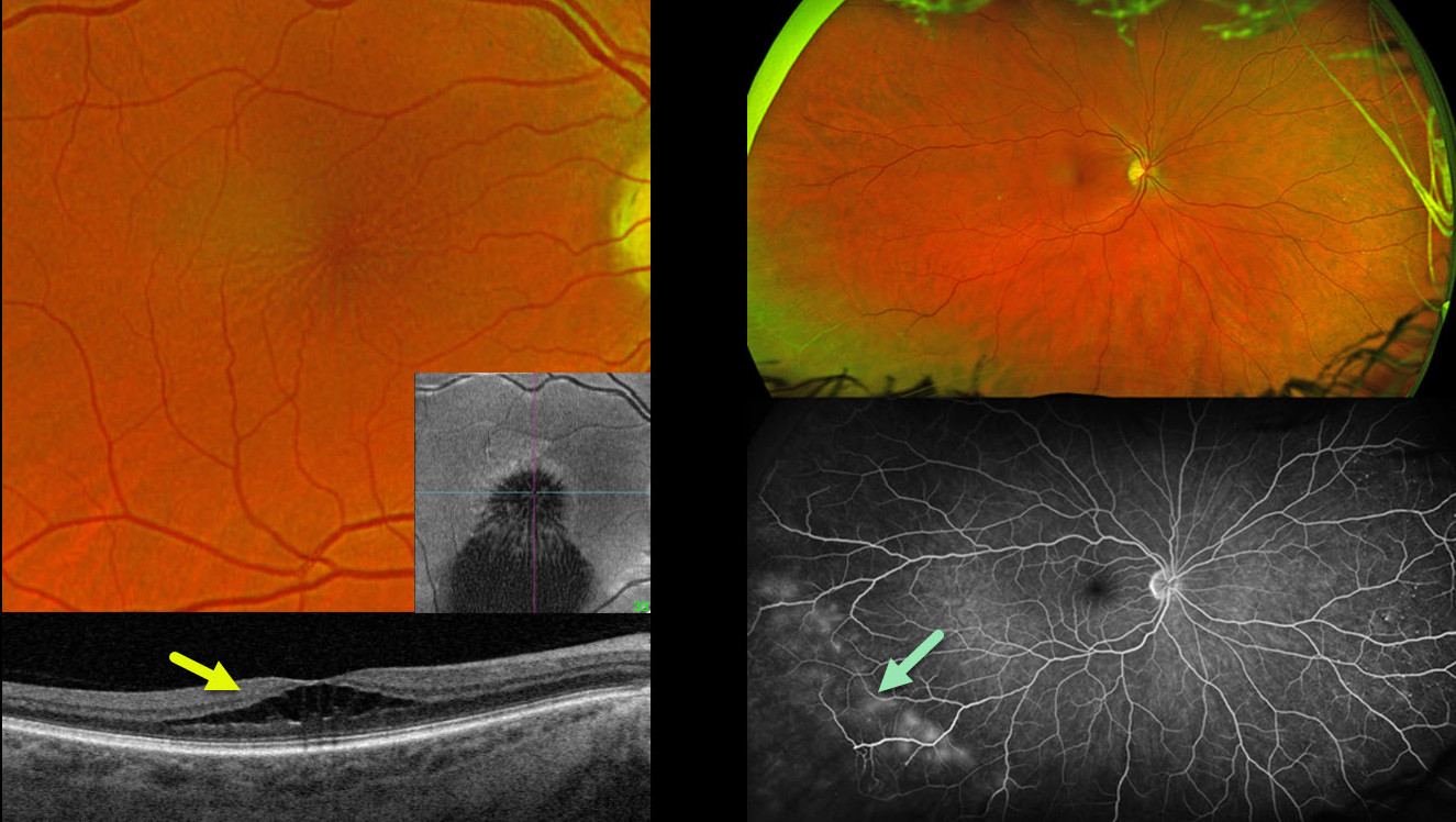
This 58YO female presented with 6 months of blurred vision in her right eye. Vision was 20/40 OD and 20/25 OS.
Optos color RG imaging of her right eye shows a stellate pattern of foveal schisis, which is confirmed on Triton swept-source B-scan and en face OCT. The macular appearance and OCT in her left eye were normal (not shown). Inferotemporal retinoschisis is noted OU peripherally. The peripheral schisis shows late leakage angiographically. Genetic testing was heterozygous for a pathogenic USH2A mutation.
Learning Points:
A new classification of stellate nonhereditary idiopathic foveomacular retinoschisis (SNIFR) was initially described by Ober et al (Ophthalmology 2014;121:1406-1413). Unlike X-linked schisis, which, in our experience, mostly affects the inner and outer nuclear layers bilaterally, SNIFR causes splitting of the outer plexiform layer, usually in just one eye. Many eyes will have peripheral retinoschisis, even when the macula is normal (Bloch et al, Retina 2021;41:2361-
X-LINKED RETINOSCHISIS
Anand Temkar and Manish Nagpal
Originally posted on @retina.rocks January 28, 2025
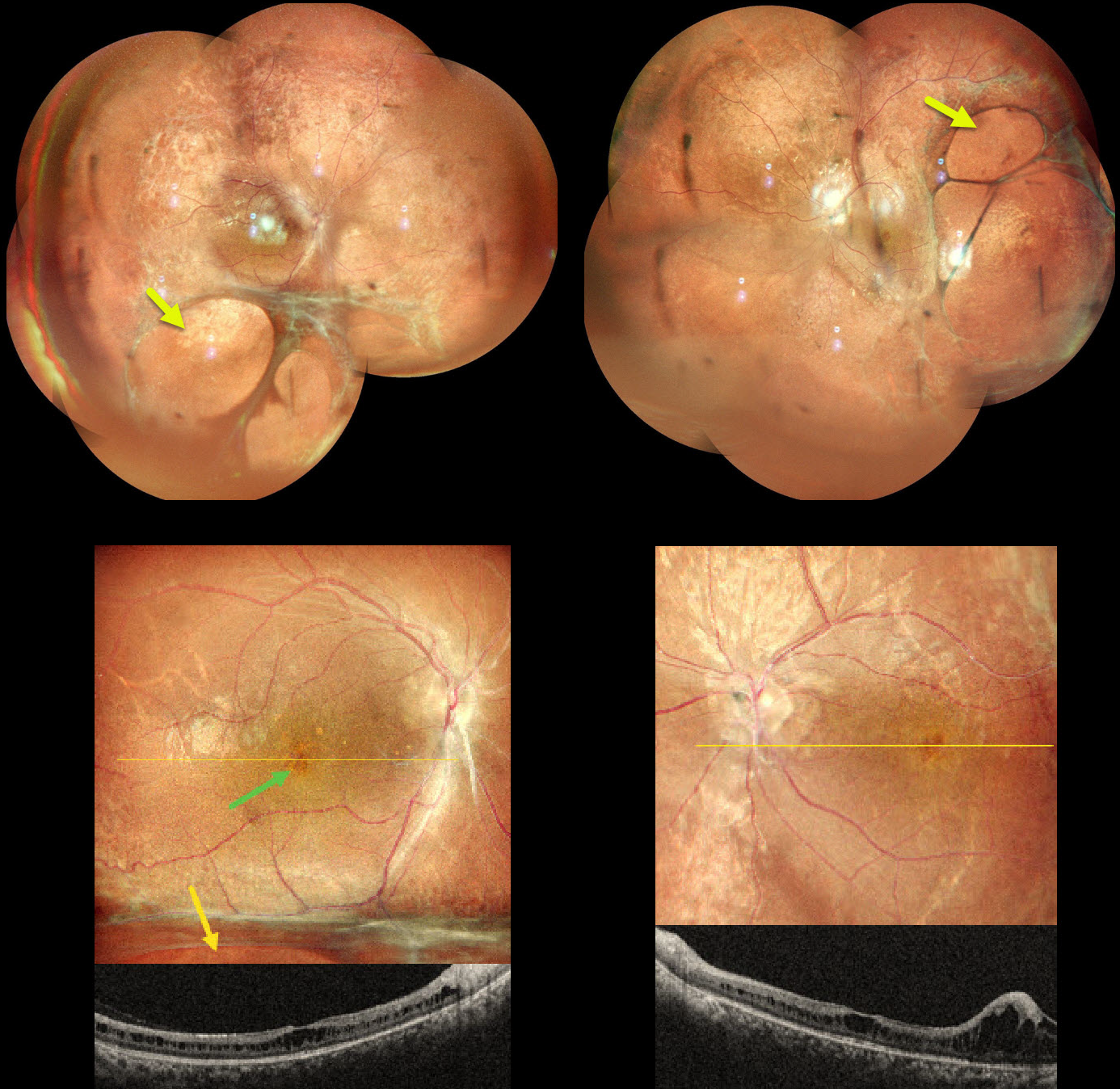
This 15YO boy presented with a lifelong history of decreased vision. His father and paternal grandfather also had poor vision but were unavailable for examination. Vision was 20/80 OD and 20/120 OS.
Pseudocolor SLO imaging shows a central, radially oriented foveal schisis with an inferior schisis hole OD. OCT scanning confirms foveal schisis, which primarily involves the inner nuclear layer. Larger areas of inner schisis extend inferiorly OD and temporally OS.
Learning Points:
X-linked retinoschisis is caused by a mutation of the RS1 gene, which is located on the X chromosome and encodes for retinoschisin, a protein secreted by photoreceptors that is involved in intercellular adhesion and likely retinal cellular organization (Heymann et al, Progress Retinal Eye Research 2023;95:101147). Although usually transmitted as an X-linked recessive disorder in males with an incidence of 1 in 15,000 to 30,000, it can also sometimes affect females.
The characteristic features include foveoschisis and peripheral schisis with vitreous veils. Over time, the macular schisis flattens with secondary atrophy. Vision usually levels off at about 20/100. Patients can develop spontaneous vitreous hemorrhage and rhegmatogenous retinal detachment from peripheral schisis. Although there is no specific treatment, topical carbonic anhydrase inhibitors may decrease the foveal thickness and possibly help minimize foveal atrophy (see Andreuzzi et al Retina 2017;37:1555-1561).
X-LINKED RETINOSCHISIS
Kanwaljeet Harjot Maden
Originally posted on @retina.rocks December 17, 2024
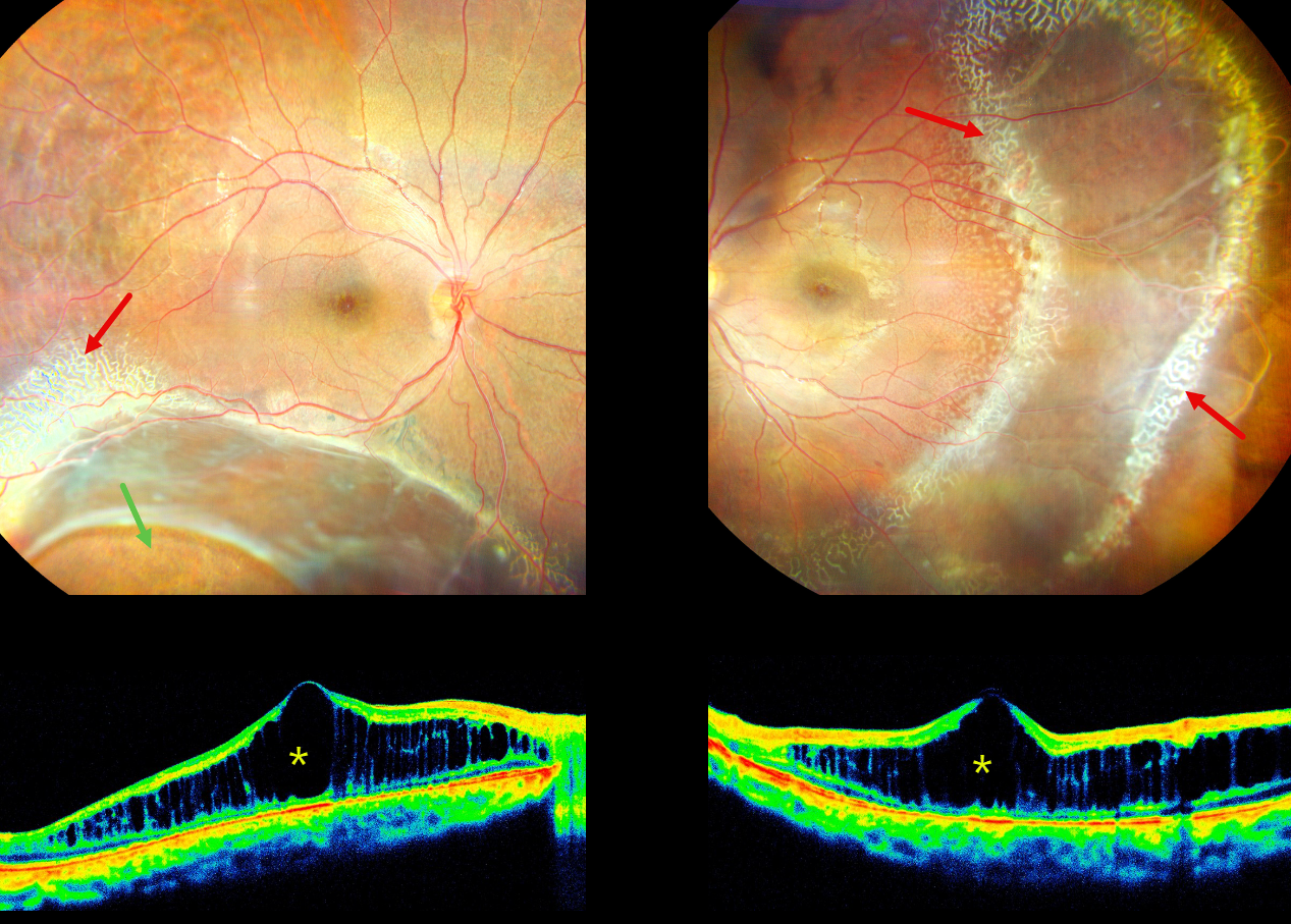
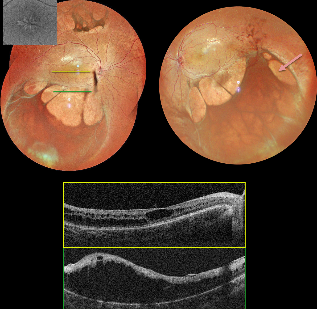
This 16YO male presented with stable bilateral vision loss since childhood, with new floaters in his right eye for 2 weeks. There was no ocular family history. Vision was 20/200 OD and 20/60 OS.
His right eye shows a large area of inferotemporal combined retinoschisis-rhegmatogenous retinal detachment (RD) extending up to the inferotemporal arcade. Subretinal pigmentation is seen along the posterior edge of the schisis inferiorly, and a large oval inner retinal defect is present inferotemporally. A combined schisis-RD is noted in his left eye which extends temporally and inferiorly. Radiating lines of foveal schisis are present bilaterally.
A dendriform pattern of occluded vessels is noted along the superotemporal edge of the schisis OD and surrounding the schisis OS. There are also numerous tiny round inner retinal breaks posterior to these dendriform lesions in the distal temporal left macula. The retinal vessels are variably fibrosed overlying the schisis cavities. OCT scanning shows severe macular schisis, primarily affecting the inner nuclear layer. Bilateral laser demaration was performed along the posterior edges of the schisis-retinal detachments.
Learning Points:
X-linked retinoschisis is caused by a mutation of the RS1 gene which is located on the X chromosome and encodes for retinoschisin, a protein secreted by photoreceptors which is involved in intercellular adhesion and likely retinal cellular organization (Heymann et al, Progress Retinal Eye Research 2023;95:101147). Although usually transmitted as an X-linked recessive disorder in males with an incidence of 1 in 15,000 to 30,000, it can also sometimes affect females.
X-LINKED RETINOSCHISIS
Tejaswita Verma and Manish Nagpal
Originally posted on @retina.rocks October 10, 2024
This 17YO boy presented with a few days of mild bilateral blurred vision. Vision was 20/40 OU.
MultiColor imaging shows central radiating foveal schisis, and larger areas of schisis extend beyond the arcades. OCT scanning confirms foveal schisis and more peripheral macular schisis. Identical findings were noted in the left macula (not shown).
Learning Points:
Sex-linked retinoschisis is caused by a mutation of the RS1 gene which is located on the X chromosome and encodes for retinoschisin, a protein secreted by photoreceptors that is involved in intercellular adhesion and likely retinal cellular organization (Heymann et al, Progress Retinal Eye Research 2023;95:101147). Although usually transmitted as an X-linked recessive disorder in males with an incidence of 1 in 15,000 to 30,000, it can also sometimes affect females.
The characteristic features include foveoschisis and peripheral schisis with vitreous veils. Over time, the macular schisis flattens with secondary atrophy, and vision usually levels off at about 20/100. Patients can develop spontaneous vitreous hemorrhage and rhegmatogenous retinal detachment from peripheral schisis. Although there is no specific treatment, topical carbonic anhydrase inhibitors may decrease the foveal thickness and possibly help minimize foveal atrophy (see Andreuzzi et al Retina 2017;37:1555-1561).
CRB1-RELATED FOVEOSCHISIS
Mattie Adams
Originally posted on @retina.rocks September 19, 2023
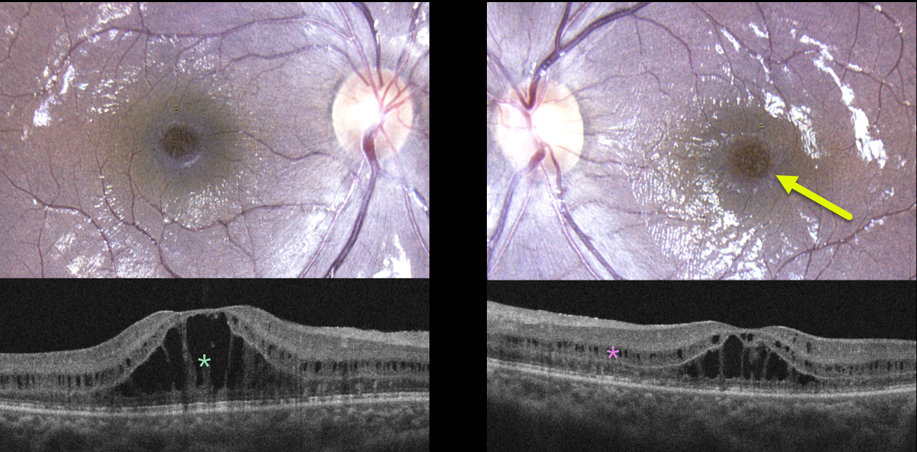
This healthy 12YO girl was referred for bilateral macular changes. She was having some mild subjective central vision loss. Vision was 20/40 OD and 20/30 OS. There was no ocular family history.
Color photography shows symmetrical radiating foveoschisis. OCT scanning shows central outer nuclear layer schisis with milder, more peripheral inner nuclear layer schisis.
Learning Points:
X-linked retinoschisis is caused by a mutation of the RS1 gene, which is located on the X chromosome and encodes for retinoschisin, a protein likely involved in retinal cellular adhesion (Heymann et al, Progress Retinal Eye Research 2023;95;101147).
Although usually transmitted as an X-linked recessive disorder found in males, non-sex-linked foveoschisis is present in other heritable disorders, such as enhanced S-cone syndrome and autosomal recessive bestrophinopathy, or non-heritable disorders, such as stellate nonhereditary idiopathic foveomacular retinoschisis.
Genetic testing of our patient revealed two pathogenic mutations of CRB1. Although few cases have been described (Vincent et al, Invest Ophthalmol Vis Sci 2016;57;2637-2646), CRB1-related foveoschisis appears to have a high female preponderance.
Similar to X-linked retinoschisis, these patients tend to maintain good vision into early adulthood, but can progress to secondary macular atrophy later in life with collapse of the schisis.
Thankfully, the foveoschisis phenotype represents the mildest of CRB1-related ocular diseases, which include Leber’s congenital amaurosis, retinitis pigmentosa, early-onset retinal dystrophy, and cone-rod dystrophy (Varela et al., AJO 2023;246:107-121).
X-LINKED RETINOSCHISIS
Originally posted on @retina.rocks April 11, 2022
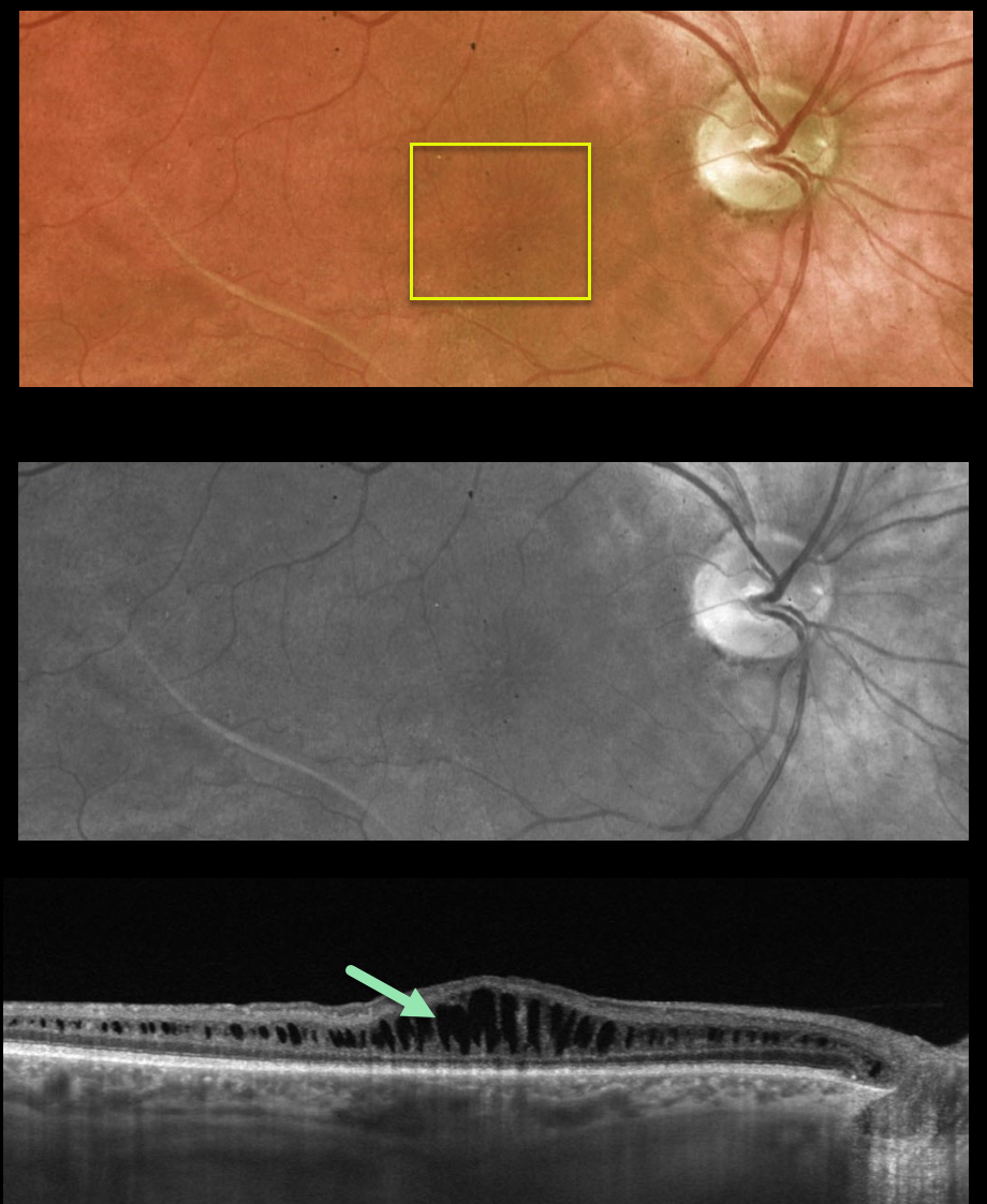
This 39YO male presented to our clinic with a known history of sex-linked retinoschisis. Vision was 20/100 OU.
Optos color and green channel images show faint radiating foveal cystic changes bilaterally (only right eye shown).
Swept-source OCT scanning shows prominent splitting of the inner nuclear layer.
Learning Points:
Although historically taught as involving the nerve fiber layer, sex-linked retinoschisis in the posterior pole causes schisis throughout the macula including the inner nuclear, outer nuclear and outer plexiform layers (see Yu et al, AJO 2010;149:973-978).
With time, the schisis often collapses with secondary atrophy (see Menke et al, Retina 2011;31:1186-1192).
X-LINKED RETINOSCHISIS
Originally posted on @retina.rocks December 10, 2021
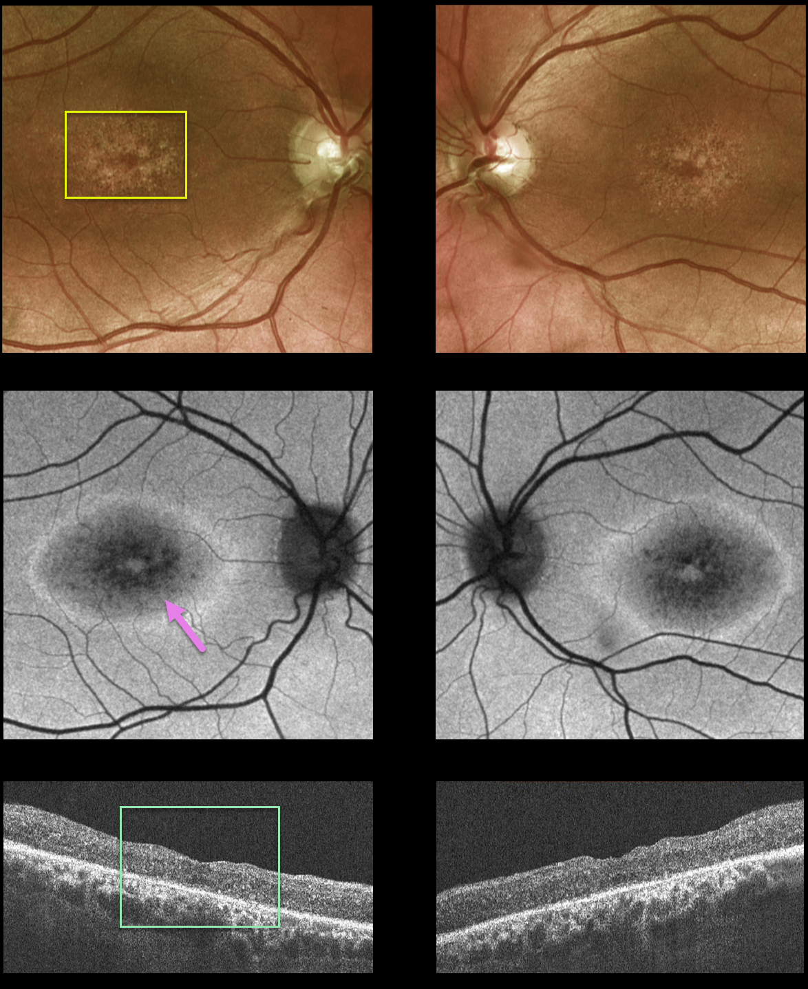
This 42YO male presented to our clinic with a history of X-linked retinoschisis diagnosed elsewhere. Vision was 20/70 OD and 20/200 OS.
Fundus photos show atrophic foveal pigmentary changes. Fundus autofluorescence (FAF) shows mottled central hypo-FAF with a ring of surrounding hyper-FAF. OCT scanning shows bilateral macular thinning with disorganization of the normal macular hyper- and hyporeflective bands.
Genetic testing confirmed the presence of a pathogenic RS1 mutation, which is associated with sex-linked retinoschisis.
Learning Points:
Although historically taught to involve the nerve fiber layer, sex-linked retinoschisis in the macula causes schisis throughout the macula, including the inner nuclear, outer nuclear, and outer plexiform layers (see Yu et al, AJO 2010;149:973-978).
Over time, the schisis often collapses with secondary atrophy, as occurred in our patient (see Menke et al, Retina 2011;31:1186-1192).
X-LINKED RETINOSCHISIS
Originally posted on @retina.rocks March 19, 2021
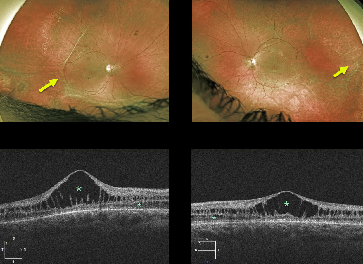
This 18YO male has classic findings for X-linked retinoschisis, including vitreous veils and bilateral foveal schisis. The macular schisis involves mostly the inner and outer nuclear layers, while peripherally it involves the nerve fiber layer (not pictured). Vision is 20/400 OD and 20/100 OS.
Learning Points:
X-linked retinoschisis is caused by a mutation in the RS1 gene, located on the X chromosome and encoding retinoschisin, a protein likely involved in retinal cell adhesion. Although it is usually transmitted as an X-linked recessive disorder in males, it can also affect females.
Over time, the macular schisis flattens with secondary atrophy. Vision usually levels off at about 20/100. Patients can develop spontaneous vitreous hemorrhage and rhegmatogenous retinal detachment from peripheral schisis.
Although there is no specific treatment, topical carbonic anhydrase inhibitors may reduce foveal thickness and possibly minimize foveal atrophy.
X-LINKED RETINOSCHISIS
Originally posted on @retina.rocks August 26, 2020
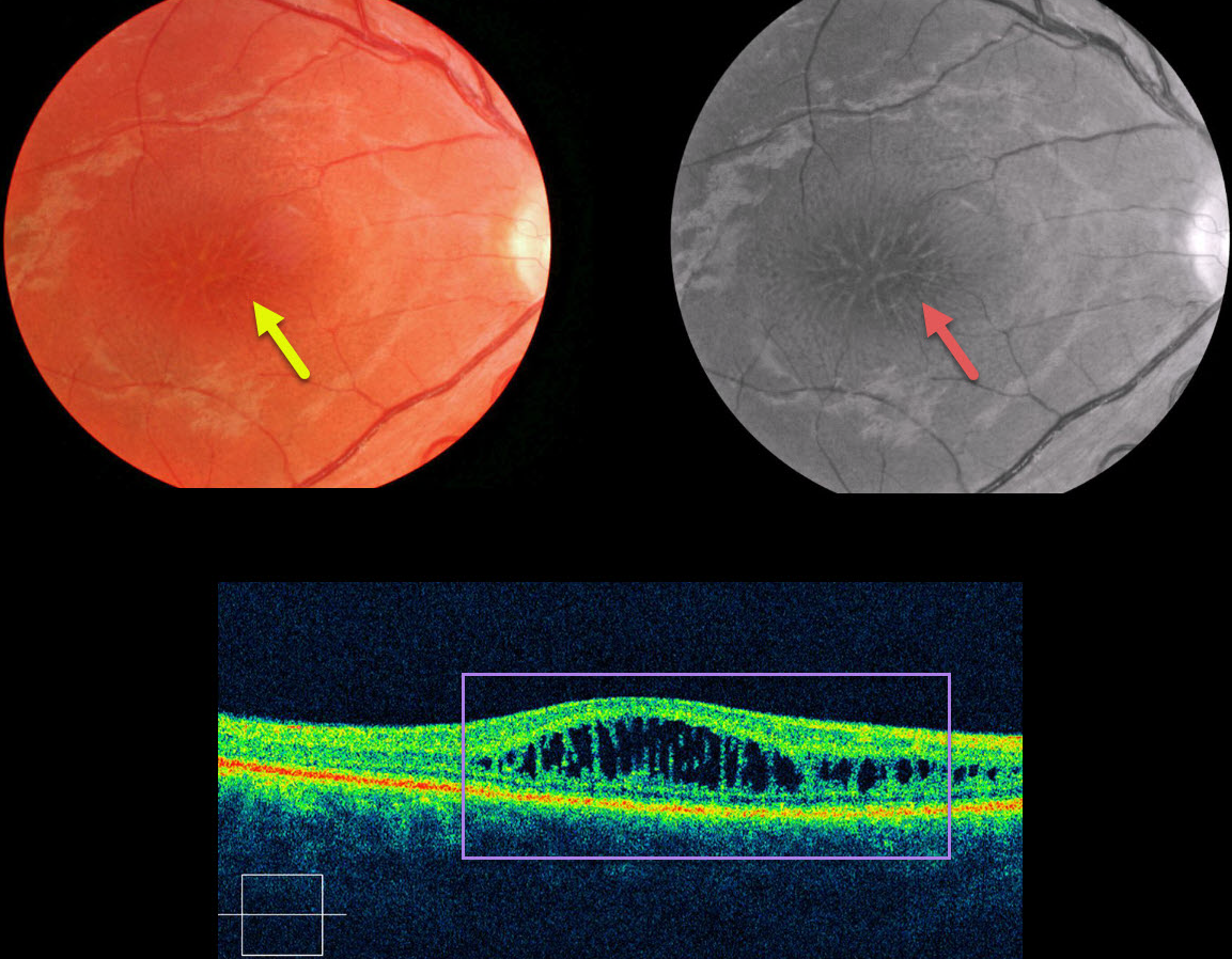
This patient presented with mild bilateral central vision loss (only the right eye is pictured).
Typical findings in sex-linked retinoschisis include radiating foveal cystoid macular edema-type changes, which are much more pronounced on red-free imaging.
Although classically described as nerve fiber layer schisis (compared to outer plexiform splitting in degenerative schisis), macular OCT here shows splitting in the inner and outer nuclear layers.
Fluoroscein angiography (not pictured) would show no leakage, as the findings are due to a structural defect rather than exudation.
Learning Points:
X-linked retinoschisis is caused by a mutation in the RS1 gene, which is located on the X chromosome and encodes retinoschisin, a protein likely involved in retinal cell adhesion. Although usually transmitted as an X-linked recessive disorder found in males, it can also sometimes affect females.
Over time, the macular schisis flattens with secondary atrophy. Vision usually levels off at about 20/100. Patients can develop spontaneous vitreous hemorrhage and rhegmatogenous retinal detachment from peripheral schisis.
Although there is no specific treatment, topical carbonic anhydrase inhibitors may reduce foveal thickness and possibly minimize foveal atrophy.
X-LINKED RETINOSCHISIS
Originally posted on @retina.rocks February 26, 2020
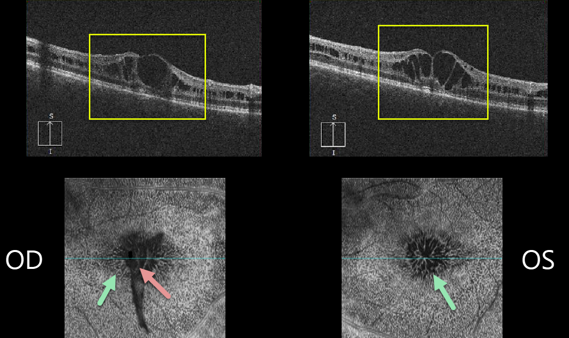
This 24YO male has bilateral 20-80 vision and foveal schisis.
B-scan OCTs show schisis mostly in the inner nuclear layer and en face OCTs show a cool perspective for this mid-retinal schisis. There is also an unusual funnel-shaped vitreous veil floating over the right fovea.
Learning Points:
Sex-linked retinoschisis is caused by a mutation of the RS1 gene which is located on the X chromosome and encodes for retinoschisin, a protein likely involved in retinal cellular adhesion.
Although usually transmitted as an X-linked recessive disorder found in males, it can also sometimes affect females.
Over time, the macular schisis flattens with secondary atrophy. Vision usually levels off at about 20/100. Patients can develop spontaneous vitreous hemorrhage and rhegmatogenous retinal detachment from peripheral schisis.
Although there is no specific treatment, topical carbonic anhydrase inhibitors may decrease the foveal thickness and possibly help minimize foveal atrophy (see Andreuzzi et al Retina 2017;37:1555-1561).


