

PENTOSAN POLYSULFATE SODIUM TOXICITY VS AMD MACULAR ATROPHY
This 59YO was examined on 9/17/19. She was taking Elmiron (pentosan polysulfate sodium, PPS) for 17 years for interstitial cystitis. Vision was 20/40 OD and 20/30 OS. Optos color RG imaging shows bilateral central-sparing macular atrophy (MA) and extensive, posterior peripheral reticular degeneration of the RPE (PRDRPE). Fundus autofluorescence shows hypo-FAF from the MA with… Read More
OPTIC PIT MACULOPATHY
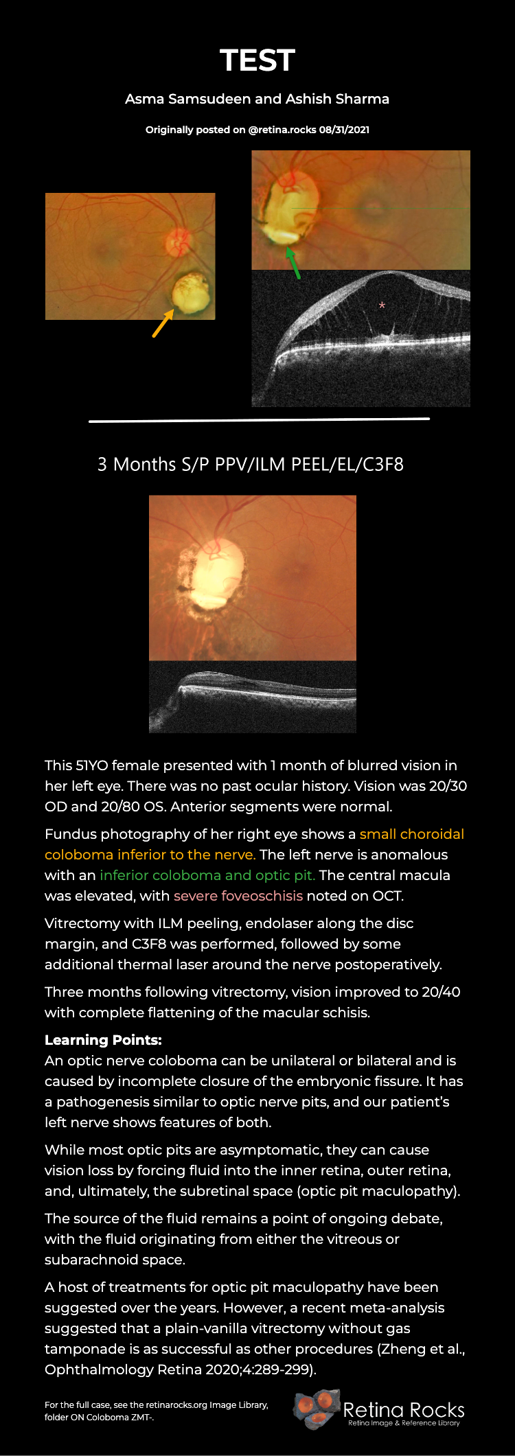
This 51YO female presented with 1 month of blurred vision in her left eye. There was no past ocular history. Vision was 20/30 OD and 20/80 OS. Anterior segments were normal. Fundus photography of her right eye shows a small choroidal coloboma inferior to the nerve. The left nerve is anomalous with an inferior coloboma… Read More
RETINITIS PIGMENTOSA + MYOPIC DEGENERATION
This 78YO female has been followed by us for several years for retinitis pigmentosa (RP) and degenerative myopia. Vision is stable at 20/30 OD and hand motion OS. Multimodal imaging shows a fascinating fusion of findings for these two disorders. Optos color RGB imaging shows typical RP findings of bone spicules (intraretinal pigment migration) extending… Read More
DIABETIC MACULAR EDEMA (DME)
This 57YO man presented on 5/26/22 with asymptomatic diabetic macular edema (DME) in his left eye. Vision was 20/20. Extensive lipid exudates with associated macular thickening were noted in the temporal macula. A gentle thermal laser was applied to the leaking vessels. One year later, vision remains at 20/20, with total resolution of all lipid… Read More
SOLAR RETINOPATHY
This 67YO female was referred for 3 weeks of vision loss in her left eye. Vision was 20/25 bilaterally. The fundus photo is normal, but her OCT shows a focal area of hyperreflectivity interrupting the ellipsoid zone. On further questioning, her symptoms developed while driving, immediately following a transformer explosion. Learning Points: The outer retinal… Read More
TOXOPLASMOSIS
This 22YO male presented with several days of central blurred vision in his left eye. There was no prior ocular or medical history. Vision was 20/25 in his normal right eye and 20/25 in his left eye. Color photography shows a variably pigmented comma-shaped colobomatous scar in the superior macula. A tiny depigmented scar is… Read More
PROLIFERATIVE VITREORETINOPATHY (PVR)
This 76YO male presented with a vague history of recent vision loss in his right eye. Vision was counting fingers. Optos color RGB imaging shows a total rhegmatogenous retinal detachment (RRD) with diffuse posterior proliferative vitreoretinopathy (PVR). The detachment was successfully repaired with vitrectomy and silicone oil. Learning Points: PVR, which consists of cellular membranes… Read More
LIPEMIA RETINALIS
This 47YO female presented with bilateral symmetric creamy colored blood vessels. Her lipid panel results were: Cholesterol 1039, Triglyceride 821, HDL 7, and LDL too high to measure. She was immediately referred for medical management of her previously undiagnosed hyperlipidemia. Learning Points: Lipemia retinalis is a rare retinal manifestation of severe hypertriglyceridemia, characterized by creamy… Read More
VALSALVA RETINOPATHY
This 37YO male presented 1 day after sudden vision loss in his right eye following vigorous coughing. Vision was 20/400 in his right eye and 20/20 in his normal left eye. Fundus examination of his right eye showed scattered posterior pole retinal hemorrhages and a large sub-internal limiting membrane (ILM) hemorrhage obscuring his right macula…. Read More
RETINITIS PIGMENTOSA WITH CHOROIDAL CAVITATIONS
This 32YO female presented with a known history of severe vision loss from retinitis pigmentosa (RP). Her parents are first-degree cousins, and there was no family history of RP. Vision was hand motion OD and counting fingers OS. Fundus photography shows bilateral irregular yellow central macular pigmentary changes, with a central hyperpigmented spot OS. OCT… Read More


